DEC Online Database
![]()
About the Online Database
| DEC OMERO Server | ||||||||||||||||
| ||||||||||||||||
The collection scanned images are currently available as a test site with only a few sample images loaded. There is no unregistered user access available to the server. All users must apply for an account and agree to abide by our access and reuse conditions before being issued a user account. Individual user accounts currently only have access to specific folders. Access to other collection folders can be managed through user rights management.
Collections have been organised into folders named after their location city, as shown below.
- Amsterdam
- Barcelona
- Berlin
- Bochum
- Göttingen
- Kyoto
- Madrid
- Newcastle
- Sydney
- Washington
2017 Test Image Link
This only in a trial, final database will vary from this draft structure.
- Folder - Each embryo has own folder (with the original embryo name as it exists in your collection).
- Project Details - CRL in mm and total number of slides available (from catalogue).
- Tag - Plane of embryo section (Sagittal, Frontal, Transverse).
OMERO Training Manual
20 July 2017 - Please note error on Page 4 of training manual the server address to enter should be 149.171.80.223 not the full web address shown in the original manual (http://149.171.80.223:8080). My apologies for this error.
Please read the DEC OMERO training manual before uploading images to the test version of the image server.
July
Locally uploaded image samples.
- Blechschmidt Collection in Christop Viebahn folder http://149.171.80.223:8080/webclient/?show=dataset-641
- Hinrichsen Collection in Beate Brand-Saberi folder http://149.171.80.223:8080/webclient/?show=dataset-735
- Hubrecht Collection in Peter Giere folder http://149.171.80.223:8080/webclient/?show=dataset-786
June
Selected slides form the Blechschmidt Collection are available for registered user access. Please advise me if you are unable to see the linked files below following log-in.
- 2.57 mm sagittal - 22 slides total 23 (1954-08-10)
- 3.4 mm sagittal - 82 slides total 87 (1954-04-09)
- 4 mm sagittal - 3 slides total 4 (1951-05-05)
- 5.8 mm frontal - 54 slides total 77 (1954-09-20)
- 6.5 mm sagittal - 40 slides total 40 (1953-09-29)
- 11 mm frontal - 29 slides total 76 (1951-09-01)
2016 Test Image Link
Note - You will need to login to access the original image through the links, images appearing on this current page are only screenshots.
Sydney folder -> 2010-08-29-1 -> Stage 22 Human Embryo (Mark Hill-001.svs) dimensions 32538 x 28770
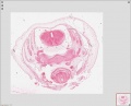
Full Image
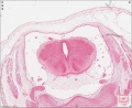
Spinal Cord

Spinal cord central canal


Image Formats
The original decision was that scanned slides would be saved in JPGXR format as these are substantially smaller than the uncompressed format. Note that OMERO server will accept embryology images in a variety of formats, including uncompressed images, the problem will be the overall size of the database would be unmanageable.
Following collection scanning, each collection manager should also hold an uncompressed archive of their own images. Should you require images of this quality/resolution, please contact the original collection managers.
CZI
The Axioskan slide scanner generates images in the CZI file format that was developed by ZEISS to specifically meet the requirements of imaging in microscopy. This is an excellent uncompressed research image format, but generates images that would be far too large for easy access on the DEC image database.
JPG XR
The Axioskan slide scanner software can also save or convert images into the JPG XR format. Note that the image file extension will still be shown as CZI, but this will be a "compressed" file. This format was selected as it has not licensing requirements, generates smaller good quality images, and the scanner can readily batch generate this format.
The JPG XR file format is a still-image compression standard and format for continuous tone photographic images, based on technology originally developed and patented by Microsoft under the name HD Photo (formerly Windows Media Photo). It supports both lossy and lossless compression, and is the preferred image format for Ecma-388 Open XML Paper Specification documents (Wikipedia). This file format is also an International Organization for Standardization ISO/IEC TR 29199-1:2011.
There are several research conversion programs, including Fiji (ImageJ), that can both read and generate this image format.
Additional non-research programs are available for image conversion into this form (Google search JPGXR conversion).
- Caution - Users should be extremely careful when downloading software from the internet and should contact their local IT managers and use anti-virus software to screen before opening, installing or running any such downloaded program.
JPEG 2000
OMERO Bioformats should also be compatible with the [ JPEG200 image format, though we have had some initial issues with this on the database. For example the image will upload to the database, but will not be able to generate the thumbnail or process the required pyramid.
- Problem - One Image 3 formats only the JPG version has generated a thumbnail image, both the TIF and J2K versions show that they are still "generating a pyramid".
- Problem - J2K image similar problem.
- OK - JPF image photograph not a scanner image uploaded and displayed without a problem. (see JPF extension)
JPEG 2000 is both a codec and a file format and the standard is in many parts.
- Part 1 giving (mostly) codec information (how to compress/decompress image data), with a container file format annex (JP2).
- Part 2 gives many extensions, and a more comprehensive container format (JPX).
Apply for DEC Account
For user account requests, please contact Dr Mark Hill.
Terms of Use
By using the DEC OMERO Server, you agree to the following:
- You agree to the specific conditions set by each collections terms of image reuse, publication and copyright.
- Reuse of images from multiple collections requires approval from each collection.
- You use this service at your own risk, and we provide NO GUARANTEE OR WARRANTY WHATSOEVER regarding uptime, security, data provenance or access, or any other service.
- You should not use the DEC OMERO Server as a repository for any important data, or data that must be held in any secure or specific way.
- The responsibility for any data loaded onto the DEC OMERO Server is entirely your own.
- We reserve the right to remove any images, data, accounts or other data or structure without notice or request.
- We welcome comments or ideas from your use of and experience with the DEC OMERO Server.
About OMERO
OMERO is client-server software for visualization, management and analysis of biological microscope images. © 2000-2016 University of Dundee & Open Microscopy Environment. Creative Commons Attribution 4.0 International License OME source code is available under the GNU General public license or through commercial license from Glencoe Software.
Version - OMERO.web 5.0.8-ice35.
References
<pubmed>20513764</pubmed>
<pubmed>22373911</pubmed>
Links
- The Open Microscopy Environment
- OMERO 5.4 Download
- OME Help
- Using OMERO.web
- Training Course Manual
- Importing Data with OMERO.insight Version 5
- JCB DataViewer
- Fiji (ImageJ)
- Image formats
Main Page | Embryo Collections | Slide Scanning | Image Server | News | Links | Test page | Site Map